Authors: Sam Engrav, Amanda Saunders*, John Walsh* MD MHPE
Background
A lower gastrointestinal (GI) bleed is any GI hemorrhage that occurs after the ligament of Treitz (suspensory ligament of the duodenum) that marks the duodenojejunal junction. Most lower GI bleeds take place in the colon, but small bowel bleeds are often more obscure, and can lead to increased interventions (imaging, transfusions, etc.) and potential complications.
 Image: Ligament of Treitz in relation to the duodenum. (Image credit: Bhalla V.P et al.).
The origin of a lower GI bleed can be divided into the following etiologies:
- Vascular
- Angiodysplasia
- Diverticulosis
- Hemorrhoids, anal fissures
- Ischemia
- Post procedure/treatment (biopsy, polypectomy, radiation)
- Inflammatory
- Infection (Campylobacter, E. coli, Salmonella, Shigella, Yersinia, etc.)
- Inflammatory Bowel Disease (Crohn’s Disease, Ulcerative Colitis)
- Ulcer
- Neoplastic
- Polyp
- Tumor – Adenocarcinoma (most common in the colon), carcinoid tumors, lymphomas, sarcomas, neuroendocrine tumors (more likely to be present in the small intestine)
- Gastrointestinal stromal tumors (GIST’s) are more likely to be present in the stomach, but rarely can manifest in the small intestine and cause bleeding
- Other
- Coagulopathies
Diverticulosis is the most common cause of a lower GI bleed, followed by various forms of colitis (infectious, inflammatory, idiopathic). In cases of diverticular bleeding, patients typically present with painless bleeding per rectum. The majority of these cases resolve spontaneously, but severe cases may require diagnosis and treatment via colonoscopy or radionuclide imaging. Treatment in these cases includes selective embolization, vasopressin infusion, or surgery. Documentation of medical intervention is important.
Similar to upper GI bleeds, there will be factors (clinical history, medications, imaging) in a decedent’s medical record that indicate a possible cause for a lower GI bleed, or at least, narrows the differential.
Quick Tips at Time of Autopsy
Clinical History
The clinical history is crucial in narrowing down the differential of a lower GI bleed. Evaluating the decedent’s demographic information, past medical history, labs, imaging, and medications should be done prior to commencing the autopsy to understand the condition of the patient and any interventions performed.
- Demographics
- In patients >65 years old, consider angiodysplasia, diverticulosis, and/or ischemic colitis
- In patients <40 years old, consider anorectal etiologies (anal fissures, hemorrhoids), and infectious/inflammatory etiologies, colorectal cancer.
- Pediatric specific considerations:
- Hirschsprung-associated enterocolitis
- Intussusception
- Malrotation with midgut volvulus
- Meckel’s diverticulum
- Necrotizing enterocolitis
- Past medical history
- Symptoms of a lower GI bleed:
- Bleeding
- Hematochezia (passage of bright red or maroon blood/stool per rectum) is typically indicative of a lower GI bleed, specifically in the colon or rectum
- Melena (passage of dark, tarry stools containing digested RBCs) mostly originates from an upper GI bleed, but can also be due to a small intestine or right colon lower GI bleed
- Abdominal pain
- Painless bleeding points towards diverticulosis
- Abdominal pain can indicate an infectious/inflammatory etiology or malignancy
- Constitutional symptoms – infection, malignancy
- Unintentional weight loss, fatigue, cachexia
- Previous lower (or upper) GI bleeds, and cause (if known)
- History of any bleeding disorders
- History of GI malignancy
- Personal or family history of inflammatory bowel disease (Crohn’s disease, ulcerative colitis)
- Underlying risks for ischemic events (or a precipitating ischemic event)
- Hypotension
- Heart/kidney failure
- Arrhythmias
- History of any lower GI intervention
- Biopsy/polypectomy (as late as 3 weeks post procedure)
- Radiation telangiectasia/proctitis
- Acute – within 6 weeks
- Chronic – 9 – 14 months after radiation (occasionally years)
- Medications
- Risk for ulcer induction:
- NSAIDs (even selective COX-2 inhibitors, like celecoxib)
- Bisphosphonates
- Antibiotics – various antibiotics can lead to increased risk for infectious colitis (and/or be used to treat it)
- Ampicillin, clindamycin, and cephalosporins increase the risk for C. diff which can lead to pseudomembranous colitis, a form of hemorrhagic colitis.
- Anticoagulants/antiplatelets
- Lab results
- Relevant labs may include a complete blood count (CBC), electrolyte panel, liver function tests
- Coagulation studies may be present if a patient has a coagulopathy or medications predisposing them to such
- Stool samples may have been taken if the patient was being worked up for an infectious colitis
- Imaging/Results
- Colonoscopy is the gold standard for diagnosis of a lower GI bleed
- CT (+/- angiography) may also be used to evaluate for malignancy, or if colonoscopy is normal despite lower GI bleeding
- Also helpful to evaluate extra intestinal lesions
- If the colon/rectum is not the source of a suspected lower GI bleed, enteroscopy may have been used to evaluate the small bowel
- In some cases, explorative surgery may have been used to evaluate a potential bleed – intra-operative notes may be useful
External examination
- External examination is more likely to yield signs of underlying/comorbid diseases.
- Examine the decedent for pallor as well as pale mucous membranes
- Examine the rectum for blood and take note of color and consistency
Internal examination
- Evaluate the GI tract in situ to check for any overt discoloration, lesions, points of hemorrhage, and/or hypoxic tissue
- Mild ischemia may look like normal postmortem changes, while significant ischemia will appear as dusky blue/darkening mucosa with potential bowel dilation
- Smell will be a potent indicator of significant ischemia
- Lesion sizes should be documented, as well as location – distance from the ligament of Treitz or the anorectal region
- Ensure if there is free blood in the abdomen this is quantified and documented even if the source is not obvious.
- Ulcers may be superficial or perforating (more likely to be present in the small intestine)
- Mild ischemia may look like normal postmortem changes, while significant ischemia will appear as dusky blue/darkening mucosa with potential bowel dilation
Opening of the GI Tract:
- Inflammatory colitis (Crohn’s disease, ulcerative colitis) present in more distinct patterns
- Crohn’s disease: segmental “skip” lesions, mucosal ulcers forming a “cobblestoning” pattern, in the small intestine and colon
- Fistulas may also form
- Ulcerative colitis: continuous stretches of superficial ulcer formation (and potentially pseudopolyps), confined to the colon

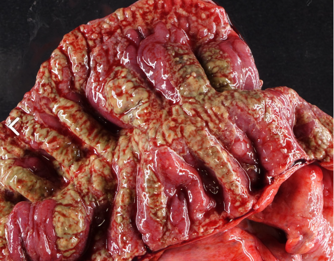
 Image: (Left) Gross appearance of ischemic bowel with dilated lumen and pseudomembranes (yellow-green exudate). (Right) Ischemic bowel with discoloration and mucosal edema compared to the uninvolved portion on the left. (Image credit: PathologyOutlines)
 Image: Gross image of the terminal ileum with irregular nodularity (green circle), hyperemia, and focal ulceration (black circle), consistent with Crohn’s disease. (Image credit: WebPath)
 Image: Gross image of the colon with a stretch of continuous inflammation from the sigmoid colon through the ascending colon. (Image credit: WebPath)
- While diverticulitis is predominantly left-sided, diverticulosis (and resultant bleeding) is predominantly right-sided in the colon
- Diverticula should be longitudinally opened and sampled
 Image: Gross image of the colon with multiple, non-inflamed diverticula with openings to the lumen of the colon. (Image credit: WebPath)
- Of note, 15-30% of patients suspected of having a lower GI bleed actually have an upper GI bleed
Quick Tips at Time of Histology Evaluation
- Depending on the etiology of a lower GI bleed, there may be findings indicating a probable etiology, or nonspecific findings
- Nonspecific signs of ischemia may be present in tissues. Early changes may include loss of surface crypts and regenerative atypia, while later changes show inflammation, crypt distortion, and hemosiderin-laden macrophages.
- Ulcers should be evaluated for size and depth, but will likely only show nonspecific signs of inflammation and/or healing
- Ulceration patterns should also be noted (if present) to indicate a potential etiology of colitis
- Chronic ulceration is useful in indicating inflammatory bowel disease
- Crohn’s disease – transmural inflammation
- Ulcerative colitis – inflammation limited to the mucosa
- Chronic ulceration is useful in indicating inflammatory bowel disease
- Ulceration patterns should also be noted (if present) to indicate a potential etiology of colitis
 Image: H&E stain of the small intestine with transmural inflammation and fissure formation though the mucosa and submucosa, consistent with Crohn’s disease. (Image credit: WebPath)
![]() Image: Low power H&E stain of the colon with transmural inflammation extending through the muscularis to the serosa adjacent to fat, consistent with Crohn’s disease. (Image credit: WebPath)
Image: Low power H&E stain of the colon with transmural inflammation extending through the muscularis to the serosa adjacent to fat, consistent with Crohn’s disease. (Image credit: WebPath)
 Image: Low power H&E stain of the colon with inflammation limited to the mucosal layer and concomitant “flask-shaped” ulcerations consistent with Ulcerative colitis. (Image credit: WebPath)
 Image: (Left) High power H&E stain of the colon with chronic ulcerative colitis showing significant mucosal inflammation with loss of goblet cells (green circle) and surface exudate (black circle). (Right) High power H&E stain of the colon with chronic ulcerative colitis showing crypt abscesses. (Image credit: WebPath)
- Mucosa of diverticula can range from normal to significantly abnormal. Findings may include:
- Expansion of lamina propria, extension of the muscularis mucosa in crypts
- Lamina propria fibrosis
- Crypt elongation
- In cases of acute diverticulitis – may also see lymphoid aggregates, cryptitis, and/or abscesses (in the crypts of around the diverticula)
 Image: A low power H&E stain of a colonic diverticulum showing a central lumen with surrounding mucosa without muscularis involvement in the diverticulum (Image credit: WebPath)
- Hemorrhoid sampling may be done if indicated
- Typically will show dilated and/or thin vascular plexus and fibrosis
- Erosion, inflammation, thromboses, and/or metaplastic changes may be present
Quick Tips at Time of Reporting
- Etiology of the bleed is crucial to report (if possible)
- If etiology is unknown after completion of autopsy, can comment on histologic findings
- Example cause of death statement:
- Gastrointestinal hemorrhage
- due to invasive adenocarcinoma of the colon.
- Hypovolemic shock
- due to Sigmoid colon hemorrhage
- due to Diverticulosis, complicated by warfarin use.
- Gastrointestinal hemorrhage
References
- Amin SK., Antunes C. Lower Gastrointestinal Bleeding. Updated 2023. Treasure Island (FL): StatPears Publishing; 2025. Lower Gastrointestinal Bleeding – StatPearls – NCBI Bookshelf
- Challa B., Yearsley MM. Diverticulosis. Updated 2022. Pathology Outlines. Diverticulosis – Pathology Outlines
- Ghassemi KA., Jensen DM. Lower GI bleeding: epidemiology and management. Curr Gastroenterol Rep. 2013; 15(7): 333. doi: 10.1007/s11894-013-0333-5.
- Gunjan D., Sharma V., Rana SS., and Bhasin BK. Small bowel bleeding: a comprehensive review. Gastroenterol Rep (Oxf). 2014; 2(4): 262-275. doi: 10/1093/gastro/gou025.
- Horton R., Hagen CE. Ulcerative colitis. Updated 2024. Pathology Outlines. Ulcerative Colitis – Pathology Outlines
- Patel N. and Kay M. Lower gastrointestinal bleeding in children: Causes and diagnostic approach. Updated 2024. Lower gastrointestinal bleeding in children: Causes and diagnostic approach – UptoDate
- Patel R., Hueller J. Hemorrhoids. Updated 2023. Pathology Outlines. Hemorrhoids – Pathology Outlines
- Ricuarte AL., Hagen CE. Crohn’s disease. Updated 2022. Pathology Outlines. Crohn’s Disease – PathologyOutlines
- Rockey DC. Lower gastrointestinal bleeding. Gastroenterology. 2006; 1:165-171. Lower Gastrointestinal Bleeding
- Strate L. Approach to acute lower gastrointestinal bleeding in adults. Updated 2025. Approach to acute lower gastrointestinal bleeding in adults – UptoDate
- Strate L. Etiology of lower gastrointestinal bleeding in adults. UptoDate. Updated 2025. Etiology of lower gastrointestinal bleeding in adults – UptoDate
* The views expressed in this article are those of the author and do not necessarily reflect the official policy or position of the Department of Navy, Naval Construction Group (NSG) TWO, Uniformed Services University of Health Science, Defense Health Agency, U.S. Navy Bureau of Medicine and Surgery, Department of Defense, or the US Government. The authors report no conflict of interest or sources of funding.


