Authors: Jesus Lopez & David S. Priemer MD
Background
Hypertension is defined as elevated pressure in the blood vessels. Traditionally coined the “silent killer,” hypertension may be asymptomatic for many years. Nevertheless, multiple organ systems are affected by chronic hypertension. This pervasive disease has far-reaching implications, given its high global prevalence, and it imposes a substantial burden on healthcare systems. Autopsy can help identify organ damage because of chronic hypertension and may play a critical role in determining the cause of death as it may relate to hypertension.
Many of the effects of chronic, benign hypertension include sequelae of atherosclerosis and arteriolosclerosis.
The American Heart Association guidelines classify hypertension as follows (current as of January 2025):
 Image: AHA guidelines on the classification of hypertension. (Image credit: AHA).
- Hypertensive urgency is classified as severe elevation in blood pressure with no evidence of acute end-organ damage.
- Hypertensive emergency is classified as severe elevation in blood pressure with evidence of acute end-organ damage.
Quick Tips at Time of Autopsy
Clinical History
Prior to the autopsy, a review of the patient’s history should include:
- When they were diagnosed with hypertension
- Medication regimen and compliance
- Known medical complications
- Evaluation for acute symptoms consistent with hypertensive crisis, including confusion, headache, chest pain, and dyspnea.
- Laboratory markers of organ damage including CK for kidneys.
- Evaluation of pre-autopsy imaging, which may show findings such as intracranial hemorrhage or pulmonary edema secondary to left-sided heart failure.
External examination
- Peripheral vascular disease/peripheral arterial disease: chronic hypertension can accelerate the process of atherosclerosis in the lower extremities; this may present as non-healing ulcers or gangrene in cases of critical limb ischemia.
 Image: Example of ulcer from peripheral artery disease. It is characterized by deep painful ulcers on the feet and ankles. In contrast, venous stasis ulcers are more superficial and tend to be found around the ankle. Diabetic foot ulcers are often painless and often occur at pressure points. Image credit: AMBOSS
- Edema in lower extremities or ascites may be indicative of heart failure, a sequela of hypertensive cardiac disease.
Internal examination
|
Internal Examination Findings in Chronic Hypertension |
||
|
Cardiovascular System |
Heart |
Concentric Left Ventricular Hypertrophy (LVH) = hypertension leads to increased work on LV which causes concentric left ventricular hypertrophy. Complications = diastolic dysfunction, ischemia, arrhythmia, sudden cardiac death
Coronary artery disease (CAD) =HTN can be associated with accelerated atherosclerosis, which narrows the coronary arteries |
|
Aorta and Large Arteries |
Gross = aortic aneurysms, dissections, and associated rupture (common in the abdomen). Complications = retroperitoneal hemorrhage secondary to rupture. | |
|
Renal System |
Nephrosclerosis |
Gross = shrunken, pitted, and granular kidneys due to changes in arterioles (arteriosclerosis) that result in ischemic glomerular sclerosis. |
|
“Malignant” Nephrosclerosis |
Associated with hypertensive crises. Gross = “flea-bitten” kidney with petechial hemorrhage. |
|
|
Renal Artery Stenosis |
Atherosclerotic changes can induce a narrowing of the renal arteries, causing stenosis and subsequent ischemic injury | |
|
Central Nervous System |
Infarcts, and Hemorrhage |
Lacunar infarct = small, non-cortical ischemic infarct related to small vessel disease including direct damage from HTN or secondary to atherosclerosis. Most commonly in the setting of benign HTN.
Intraparenchymal (classically ganglionic) HTN-related hemorrhage which can range from small to catastrophic, with possibility of ventricular extension and further possible extension into subarachnoid space via the cerebellar cisterns. Often in the setting of hypertensive crisis/emergency. |
|
Pulmonary System |
Pulmonary Venous Hypertension |
Usually a complication of left-sided heart failure secondary to the increased work of pumping blood through hypertensive arteries. |
|
Diffuse Alveolar Hemorrhage |
Associated with hypertensive emergencies, revealing diffuse alveolar hemorrhage | |
|
Ocular Findings |
Hypertensive Retinopathy |
Common findings include arteriovenous nicking, retinal hemorrhages, and cotton wool spots. In more severe cases, an edematous optic disc with ischemic changes may be noted. |


Image: Gross photograph of cardiac left ventricular hypertrophy secondary to chronic systemic hypertension. The left ventricle is concentrically thickened, and the ventricular cavity is markedly reduced in size. (Image credit: College of American Pathologists 2024 AUP-B Symposium Hypertension).

Image: Gross photograph of an aortic dissection (arrow). When due to hypertension, aortic dissections commonly originate in the ascending aorta and often extend to involve the arch and descending aorta. The dissection most commonly occurs between the inner 2/3 and outer 1/3 of the aortic wall. (Image credit: College of American Pathologists 2024 AUP-B Symposium Hypertension).

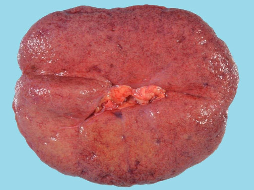
Image: Gross photograph of renal nephrosclerosis due to chronic systemic hypertension. Nephrosclerotic kidneys are often below expected weight. As opposed to being smooth and glistening, their outer surfaces are often finely granular in appearance. (Image credit: College of American Pathologists 2024 AUP-B Symposium Hypertension).
 Image: Gross photograph of lacunar infarct within the pons. Healed, gliotic cavity in the base of the pons (arrow), corresponding to a remote lacunar infarct. These small infarcts are most commonly noted in deep grey nuclei, the brainstem (pons), and cerebellar white matter in the setting of chronic systemic hypertension and resultant cerebrovascular arteriolosclerosis. (Image credit: College of American Pathologists 2024 AUP-B Symposium Hypertension).

Image: a large intraparenchymal bleed originating in the frontal hemisphere secondary to a hypertensive crisis. (Image credit: Meagan Chambers/University of Washington).
Quick Tips at Time of Histology Evaluation
- Cardiomyocyte hypertrophy and patchy myocardial fibrosis can corroborate gross findings of left ventricular hypertrophy. Notably, cardiomyocyte nucleomegaly and boxcar shape with thickened myocyte cell bodies supports hypertensive cardiomyopathy, while cardiomyocyte nucleomegaly and boxcar shape with thin/normal myocyte cell bodies supports eccentric hypertrophy/heart failure.
 Image: Cardiomyocytes with nucleomegaly (towards the upper left side of the image) are seen with normally sized nuclei (right side of the image). (Image credit: Meagan Chambers/Stanford Hospital).
- Aorta: intimal fibrosis, medial degeneration, cystic medial necrosis
- Evaluate small arterial walls for hyaline arteriolosclerosis, characterized by amorphous, eosinophilic material deposition (PAS-positive) within the vessel walls. This can be particularly prominent in kidney, and brain (subcortical structures) samples.

Image: H&E of kidney demonstrating severe arteriosclerosis characterized by thickening of the wall and eosinophilic deposits (as well as calcification in this case). (Image credit: Meagan Chambers/University of Washington)
- Nephrosclerosis: chronic HTN leads to sclerosis of the small renal arteries and arterioles which causes ischemic changes manifesting as tubular atrophy, interstitial fibrosis, and glomerulosclerosis
- Malignant nephrosclerosis: necrotizing arteriolitis characterized by fibrinoid necrosis, hyperplastic arteriolosclerosis characterized by concentric, onion-skin thickening

Image: Renal tubular atrophy and numerous globally sclerotic glomeruli in the context of severe arteriolosclerosis from chronic, benign hypertension. (Image credit: Meagan Chambers/University of Washington)
- Cerebral vessels often demonstrate lipohyalinosis, and associated Charcot-Bouchard aneurysms may be implicated in hypertensive hemorrhage.
- Cases of chronic, severe and poorly managed hypertension may be associated with subcortical arteriosclerotic encephalopathy: Also known as Binswanger disease, this is now an uncommon complication of long-standing hypertension. It is considered a form of vascular dementia, and it is pathologically characterized by widespread arteriosclerotic vascular changes, resultant brain atrophy, and often numerous multiple infarcts.
 Image: Hypertensive arteriopathy (arteriolosclerosis) in the brain, characterized by hyalinosis of the vessels. Often associated with perivascular hemosiderin deposition. (Image credit: College of American Pathologists 2024 AUP-B Symposium Hypertension).
- Pulmonary venous hypertension: edematous fluid accumulation within the alveoli and hemosiderin-laden macrophages in the setting of left-sided heart failure related to chronic hypertension (“heart-failure cells”).
Quick Tips at Time of Reporting
- Examples of cause of death statements:
- Fatal arrhythmia due to, or as a consequence of, hypertensive cardiomyopathy. Contributing conditions include chronic, benign hypertension.
- Cerebral hemispheric hemorrhage due to, or as a consequence of, systemic hypertension (hypertensive emergency).
Recommended References
- Mensah GA, Croft JB, Giles WH. The heart, kidney, and brain as target organs in hypertension. Cardiol Clin. 2002 May;20(2):225-47. doi: 10.1016/s0733-8651(02)00004-8. PMID: 12119798.
Additional References
- Buja LM, Butany J, eds. Cardiovascular Pathology, 5th edition. Elsevier Academic Press, 2022.
- Chobanian AV. Vascular effects of systemic hypertension. Am J Cardiol. 1992 Apr 30;69(13):3E-7E. doi: 10.1016/0002-9149(92)90010-v. PMID: 1575175.
- Kahan T. The importance of left ventricular hypertrophy in human hypertension. J Hypertens Suppl. 1998 Sep;16(7):S23-9. PMID: 9855028.
- Kearney PM, Whelton M, Reynolds K, Muntner P, Whelton PK, He J. Global burden of hypertension: analysis of worldwide data. Lancet. 2005;365(9455):217-223.
- NCD Risk Factor Collaboration (NCD-RisC). Worldwide trends in blood pressure from 1975 to 2015: a pooled analysis of 1479 population-based measurement studies with 19·1 million participants. Lancet. 2017 Jan 07;389(10064):37-55.
- Persu A, De Plaen JF. Recent insights in the development of organ damage caused by hypertension. Acta Cardiol. 2004 Aug;59(4):369-81. doi: 10.2143/AC.59.4.2005202. PMID: 15368798.


