Authors: Alex K Williamson MD & Meagan Chambers MD
The gross description of autopsy findings serves to record what was encountered during the postmortem examination. A written gross description should allow for its reader to visualize and recreate in his or her mind exactly what the autopsist observed during the postmortem examination; in other words, a well-composed, thoughtful, and detailed description should allow the report’s reader to draw exactly what was observed at autopsy.
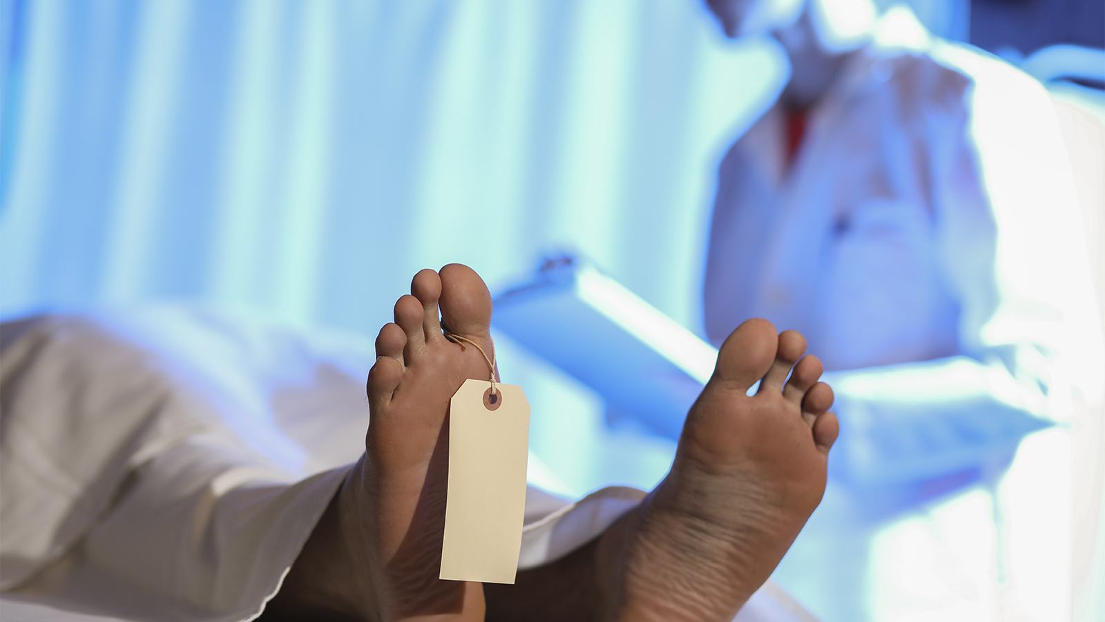
The examination begins with confirming the identify of the decedent, noting the location and contents on identification tags.
Any clothing removed from the body should be examined and documented. In hospitalized children and adults, the contents of any diaper on the body should be examined and documented.
The external examination can be performed in 3 phases, with each phase involving careful examination of the body from head to toe paying attention to the indicated evaluation each extending comprehensively from head to toe:
- Examine the native body including postmortem changes
- Evaluate interventions (e.g., tracheostomy tube, cardiovascular access lines, etc
- Document scars and other injuries (e.g., abrasions from CPR, ecchymoses from cardiovascular access, etc)
Anatomic regions of the body
- Head
- Neck
- Torso includes chest and abdomen (anterior) and back (posterior)
- Upper extremity includes the shoulder, arm, elbow, forearm, wrist, hand
- Lower extremity includes the hip, thigh, knee, lower leg, ankle, foot
- Genitalia, perineum, and groins
Anatomic terminology
Usually the body can be described with customary anatomic descriptors with the body assumed to be in a standard anatomic position (i.e., Da Vinci’s man). These terms include: superior, inferior, lateral, medial, anterior, posterior.
For the forearms, which can be in various positions in life relative to the body and standard anatomic position (i.e., arms up in defensive posture, running), better terms to describe lesions include flexor or extensor aspect and radial or ulnar aspect.
Body build
- Small frame
- Medium frame
- Large frame
Body length should always be measured at autopsy; body weight can be taken from medical record or approximated using correct body length and BMI grids with visual estimation of BMI (i.e., thin, normal, overweight, obese, morbidly obese).
Skin color
- Use objective color, not subjective race or ethnicity
- “White” or “light skinned”, “Brown”, Black” or “dark skinned”
- Avoid using “Caucasian”, “African-American”, “Asian”
If a light-skinned person is tan from sun exposure, you can describe that as “The skin is light with areas of hyperpigmentation on the head, face, extremities, and trunk consistent with sun exposure”.
Postmortem changes
- Rigor mortis: stiffening of the body after death : assessed by flexing and extending joints
- Livor mortis: gravity-dependent pooling of blood in vessels (hypostasis) after death : assessed by firmly pressing for at least 5 seconds and then releasing pressure
- Algor mortis: cooling of the body after death
Common findings
- Pitting edema should be assessed by firmly pressing for 5 seconds on affected soft body parts, such as the medial lower legs and medial thighs.
- Ecchymosis = area of discoloration; unknown etiology
Contusion = ecchymosis caused by blunt force trauma; a “bruise”
Best to use ecchymosis if unsure about etiology of lesion
- Congestion is intravascular
- Hemorrhage is extravascular blood; requires defect in vessel and circulation to defect
- Hematoma is a space-occupying collection of blood; should be measured in cm3
Abrasion = removal of superficial tissue layers (i.e., epidermis of skin or epithelium of mucosa) via blunt force trauma
- Caused by mechanical blunt force trauma applied in a perpendicular (i.e., imprinting) or parallel (i.e., scraping) direction; thermal injury can also cause removal of superficial tissue layers
Tattoos can be important in identification of identity (i.e., unknown decedent) and risk factors for disease (i.e., hepatitis virus infection). They should be described as follows:
- Professional appearing
- Polychromatic or monochromatic
- Anatomic location
- Greatest dimensions
- General description of obvious designs or words or dates
Example description: “On the lateral right shoulder there is a professional-appearing, polychromatic tattoo of a sailor design with the word “Popeye”, up to 4 in x 4 in.”
Evidence of Postmortem Tissue Harvest for Donation
- Bilateral eye tissues
- Long bones of each upper and lower extremity. Each anterior arm shows a linear incision closed with a running suture extending from the anterior shoulder to the antecubital fossa. Each medial lower extremity shows a linear incision closed with a running suture extending from the lower abdomen to the foot. Hard cylindrical pieces of material are palpated in place of the harvested bones.
Example description: “In each upper extremity there is a recent incision closed with a running suture extending from the anterior shoulder to the proximal forearm (right 37 cm, left 40 cm). In each lower extremity there is a recent incision closed with a running suture extending from the anterior hip to the medial ankle (right 95 cm, left 92 cm). Cylindrical segments of hard material are palpably present in the place of long bones underneath each of these incisions. In the torso there are two intersecting linear incisions closed with running sutures extending from the manubrium to the symphysis pubis and across the middle torso. Subsequent internal examination reveals an incision through the anterior chest wall and absent tissues and viscera including left lung, heart, aorta, liver and gallbladder, kidneys, adrenal glands, and a segment of distal small bowel into proximal colon. The residual terminal small bowel and proximal colon margins are stapled. The spleen and pancreas have each been sectioned; nearly 1/3 of the spleen and distal one half of the pancreas are absent. The diaphragm is disrupted and the remaining gastrointestinal tract has been displaced into the thorax. There is scant serosanguineous fluid in the torso.
Evidence of Therapeutic Intervention
- Orotracheal tube terminates in the trachea
- Orogastric tube terminates in the stomach
- Cardiovascular access line in right wrist.
- Foley catheter in urinary bladder and penile urethra.
- Findings consistent with cardiopulmonary resuscitation:
- A circular, brown-red, dry abrasion on the middle chest, ~5-6 cm diameter
- Multiple bilateral anterior rib fractures
- Localized hemorrhage in anterior mediastinum.
Vocabulary
- Bilateral: Doesn’t need to be used when describing a change present in both of two organs (e.g., lungs, kidneys, adrenals, upper extremities, feet, etc)
- Slight versus Mild: Mild is for temperature (weather, salsa)
- Use: Slight/Moderate/Marked
Additional Resources:


