Authors: Casey P. Schukow DO & Tanner Bartholow MD
Background
Although alcohol consumption is a common practice in many cultures, it is associated with numerous significant health risks, including multiorgan damage, use disorders/dependence, cancer, and death. Global numbers (as of 2019) on alcohol consumption are summarized below:
- Around 2.6 million deaths were attributed to alcohol consumption (1.6 million from noncommunicable diseases, 700,000 from injuries, and 300,000 from communicable diseases).
- Mortality is more common in men (2 million compared to 600,000 deaths in women).
- From 2010-2019, a 20.2% decrease in alcohol-attributed deaths per 100,000 people was seen.
The Centers for Disease Control and Prevention (CDC) defines excessive alcohol use (“drinking”) into four subcategories:
- Binge: ≥ 4 or 5 drinks (for a woman or man, respectively) in one setting (e.g., social occasion).
- Heavy: ≥ 8 or 15 drinks (for a woman or man, respectively) in one week.
Organ damage in the setting of chronic or excessive alcohol use is outlined by the National Institute on Alcohol Abuse and Alcoholism (NIAAA) and summarized below:
| Organ/System | Effects/Findings |
| Brain | Impaired communication pathways; mood/behavior swings; coordination dampening; stroke (ischemic, hemorrhagic). |
| Oral Cavity | Poor dentition (halitosis, caries). |
| Heart | Cardiomyopathies (e.g., dilated); arrhythmias; hypertension; ischemic heart disease. |
| Lungs | Acute respiratory distress syndrome; pneumonia. |
| Liver | Steatosis, steatohepatitis, fibrosis, cirrhosis, hepatitis. |
| Pancreas | Pancreatitis (acute and chronic). |
| Gastrointestinal Tract | Gastroesophageal reflux disease; gut leakiness; microbial dysbiosis. |
| Musculoskeletal System | Myopathy; muscle wasting; impaired fracture repair; reduced bone density. |
| Hematopoietic System | Immune dysregulation (chronic infections); cytopenias (anemia, thrombocytopenia, leukopenia). |
Quick Tips at Time of Autopsy
Clinical History
- Alcohol consumption often carries a stigma; reported intake may underestimate actual consumption. Objective findings are critical.
- Always consider alcohol as a contributing factor in the differential diagnosis, given its prevalence and accessibility.
- Evaluate comorbid histories (e.g., drug abuse, psychiatric conditions).
- Note a history of vitamin or essential mineral deficiencies, which are commonly associated with chronic alcohol consumption and malnutrition. Glossitis (inflamed tongue) and stomatitis (mouth inflammation) are common among most vitamin/mineral deficiencies, but the following are more specific:
- Vitamin B6 (Pyridoxine): Deficiency may contribute to sideroblastic anemia and peripheral neuropathy.
- Vitamin B9 (Folate) and B12 (Cobalamin): Deficiencies cause macrocytic anemia and neuropathies; folate is particularly affected by chronic alcohol use.
- Vitamin A: Deficiency can lead to night blindness and impaired immunity.
- Vitamin C: Bleeding gums, loose teeth, perifollicular hemorrhages (bleeding around hair follicles). Commonly referred to as “scurvy”.
- Vitamin D: Deficiency contributes to osteomalacia and impaired bone health.
- Zinc: Deficiency causes delayed wound healing, loss of taste, and acrodermatitis enteropathica-like presentations.
- Copper: Deficiency may mimic Wilson-like disease symptoms or lead to anemia and leukopenia.
- Anemia with increased MCV may also be present.
External Examination
- Acute Intoxication: No specific external markers; consider context (e.g., behavioral patterns, scene evidence, ED alcohol screening).
- Chronic Alcoholism (many of which are non-specific and require clinical correlation):
- Cachexia: Severe muscle and fat wasting.
- Jaundice: Yellowing of the skin and eyes due to elevated bilirubin. This often includes scleral icterus, a yellow discoloration of the sclera, which is an important and visible early sign of hyperbilirubinemia.
- Ascites: Abdominal swelling due to fluid accumulation, often from liver cirrhosis.
- Rosacea: Includes various forms, such as erythematotelangiectatic (redness and visible blood vessels), papulopustular (redness with acne-like breakouts), and phymatous (thickened skin with bulbous deformities, typically on the nose). Chronic alcohol use is most associated with worsening phymatous rosacea, but other forms may also occur.
- Other Skin Findings related to alcoholic cirrhosis: Chronic alcohol use may also present with palmar erythema (reddening of the palms), nail clubbing, telangiectasia/spider angiomas (spider-like blood vessels), and ecchymosis (bruising), often linked to coagulopathy or liver dysfunction.
- Self-Harm Findings: Linear scars on forearms or other body parts may suggest a history of depression, self-harm, or suicidal ideation, conditions often associated with heavy alcohol consumption.
- Vitamin/mineral deficiencies: Glossitis/stomatitis (e.g., mouth ulcers), chronic non-healing wounds, perifollicular hemorrhages.
- Comorbidities: Poor hygiene, dental decay, and systemic complications (e.g., diabetes, cardiovascular disease).
 Image: signs of vitamin C deficiency (scurvy) include bloody gums with mucosal hypertrophy (left) and folliculitis with/without corkscrew hairs (right). (Image credits: Dentist Peshwar [left], DermNet [right]).
Internal Examination
Liver
- Steatosis (Fatty Liver): Present in 90-100% of individuals with heavy alcohol use; seen macroscopically as a yellow or orangish discoloration to the liver.
- Fibrosis: Scarring of liver tissue, often progressing to cirrhosis.
- Cirrhosis: Advanced fibrosis leading to nodular liver architecture, portal hypertension, and liver failure; seen in approximately 15-20% of chronic alcoholics.
 Image: this liver in a chronic alcoholic has the typical yellowish discoloration of a steatotic liver. (Image credit: Meagan Chambers/Stanford Hospital).
Heart
- Atherosclerosis: while it is a commonly repeated anecdote in the autopsy suite that chronic, high level consumption of alcohol decreases the risk for aortic and coronary artery atherosclerosis, the literature on this area has been mixed.
 Image: This middle-aged patient with hyperlipidemia, hypertension, and diabetes (all risk factors for atherosclerosis) had only focal fatty streaking in his aorta. His cause of death was complications of long term alcohol use disorder. While this is a striking example of minimal atherosclerosis in heavy drinkers at autopsy, the association thus far is only anecdotal. (Image credit: Meagan Chambers/Stanford Hospital).
- Alcoholic Cardiomyopathy: Seen in up to 30% of heavy chronic drinkers; characterized by dilated ventricles (and reduced cardiac output on ECHO). The alcoholic cardiomyopathy cannot be distinguished from other dilated cardiomyopathies on gross examination alone.
- Hypertensive cardiomyopathy: Often co-occurring in individuals with long-term alcohol use.
 Image: This heart weighed 550 grams at autopsy and displayed a globoid shape. On sectioning there was four-chamber dilation consistent with dilated cardiomyopathy. These findings can be consistent with alcoholic cardiomyopathy (as in this patient with chronic alcohol abuse), but are not specific and are seen in other dilated cardiomyopathies. (Image credit: Meagan Chambers/Stanford Hospital).
Lungs
- Pulmonary Edema: Fluid accumulation in the lungs, often secondary to cardiac failure or aspiration.
- Aspiration Findings: Evidence of inhalation of gastric contents, more common in acute intoxication deaths.
- Infectious Associations: Chronic alcohol use predisposes individuals to specific infections by impairing immune function through multiple mechanisms, such as disrupting mucosal barriers, altering gut microbiota, and reducing the production of key immune cells (e.g., macrophages, neutrophils). This immunosuppression increases susceptibility to bacterial, viral, and fungal pathogens, with notable infections including:
- Klebsiella pneumoniae: Commonly associated with aspiration, leading to necrotizing pneumonia or abscess formation. Findings include currant jelly sputum (antemortem) and cavitary lesions.
- Mycobacterium tuberculosis: Chronic immunosuppression in alcoholics increases susceptibility. Internally, miliary nodules or caseating granulomas may be found.
- Alcoholics are more likely to have pneumococcal endocarditis.
Brain
- As many patients with chronic alcohol use have coagulopathies (secondary to liver disease), when opening the brain, the sagittal sinus can rupture and bleed. This should not be confused with a subdural hemorrhage.
- Cortical Atrophy: Shrinkage of the brain cortex, common in chronic alcoholics.
- Cerebellar Vermis Atrophy: Loss of tissue in the midline of the cerebellum, affecting coordination.
- Mammillary Body Degeneration: Classic finding in Wernicke’s encephalopathy (linked to thiamine deficiency).
- Marchiafava–Bignami disease: Chronic alcohol use can lead to necrosis and atrophy of the corpus callosum; specifically demyelination or necrosis of the central part of the corpus callosum with sparring of the peripheral fibers.
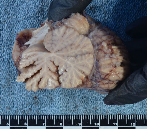
 Image: Alcoholic cerebellar vermis atrophy predominantly affects the anterior vermis (left of the image), sparing the posterior vermis. (Image credit: Meagan Chambers/University of Washington).
 Image: Macroscopic evidence of necrosis of the mammillary bodies in chronic alcohol use. (Image credit: Dr. Alvord Shaw/University of Washington).
 Image: An example of Marchiafava-Bignami disease: demyelination of the central part of the corpus callosum with sparring of the peripheral fibers as seen on LFB staining. (Image credit: Dr. John Newman/Stanford Hospital.)
Pancreas: may show features of acute and chronic pancreatitis (see the microscopic section for additional information).
 Image: This patient had chronic pancreatitis with significant loss of acini and extensive fatty replacement. (Image credit: Meagan Chambers/Stanford Hospital).
Ancillary Testing
- Tissue Cultures or other Microbiological testing: Fresh tissue samples, especially from lungs or granulomatous lesions, should be submitted for culture in suspected infectious cases (see linked article for additional information). For abscesses (e.g., Klebsiella pneumoniae), cultures can confirm the bacterial etiology. Similarly, culture of granulomas can identify Mycobacterium tuberculosis or atypical mycobacteria.
- Toxicology: may be considered in cases of acute intoxication. Post mortem specimens may include blood, vitreous humor, or urine.
Quick Tips at Time of Histology
- Brain: Gliosis, neuronal loss, and microhemorrhages may be seen with chronic alcohol use.
- Increased number of Hirano bodies have also been reported in alcoholics.
- Basal Ganglia Microhemorrhages: Small brain hemorrhages, often related to hypertension, may be seen in the setting of chronic alcohol use.
- Central Pontine Myelinolysis: Demyelination in the pons; may be due to rapid correction of hyponatremia in chronic alcoholics (as a result of low plasma sodium).

Image: Histologic examination of alcoholic atrophy of the vermis demonstrates loss of the molecular layer (right) compared to an age matched control (left). (Image credit: Meagan Chambers/University of Washington).
- Heart: Myocyte hypertrophy, interstitial fibrosis as part of alcohol-related cardiomyopathy.
- Lungs: Acute inflammation or necrosis with bacterial colonies may be present with pneumonia.
- Klebsiella pneumoniae: Necrotizing inflammation and bacterial colonies in lung abscesses.
- Mycobacterium tuberculosis: Caseating granulomas with central necrosis, epithelioid macrophages, and Langhans giant cells (can see anywhere in the body in cases of disseminated infection).
- Pancreas: Fibrosis, calcifications, and fat necrosis.
- Acute Pancreatitis: Affects approximately 5-10% of alcohol abusers; sudden inflammation with fat necrosis and edema.
- Chronic Pancreatitis: Found in 10-20% of chronic alcoholics, with fibrosis and calcifications within the gland.
- In patients with chronic pancreatitis and significant loss of acini, increased oxalate crystals may be seen in the kidneys. This can even result in acute kidney injury.
 Image: Chronic pancreatitis in a patient with chronic alcohol use. The image demonstrates significant areas of fatty replacement. Residual islands of acini and islet cells are surrounded by fibrosis. (Image credit: Meagan Chambers/Stanford Hospital).
- Kidneys: Oxalate crystals can be seen in cases of acute toxicity with ethylene glycol co-ingestion (typically these are calcium oxalate monohydrate crystals). But oxalate crystals (typically calcium oxalate dihydrate crystals) can also be seen in chronic alcohol use when there is pancreatic atrophy/chronic pancreatitis. In both cases, significant accumulation of oxalate crystals in the kidneys can result in clinically detectable AKI.
- Subnuclear vacuoles in the proximal renal tubules are classically seen in diabetic ketoacidosis. However, they are also seen in chronic alcohol use, alcoholic ketoacidosis (AKA), starvation, and even hypothermia.
 Image: Renal histology showing variations in subnuclear vacuole prominence from none (0), scanty (1), and frequent (2). (Image credit: Auld 2023).
- Liver: Macrovesicular steatosis, Mallory-Denk bodies, bridging fibrosis, siderosis, and alcoholic steatohepatitis.
- Macrosteatosis: Characterized by large fat droplets displacing the nucleus in hepatocytes; often associated with heavy alcohol consumption. Severity can be graded as mild (<33%), moderate (33-66%), or severe (>66%) based on the proportion of affected hepatocytes (detected histologically).
- First appears in perivenular region (zone 3) and spreads to other regions if drinking persists; may disappear within 1 month after alcohol cessation
- Microsteatosis: Identified by small fat droplets within hepatocytes without nuclear displacement. Often seen in conditions such as acute fatty liver of pregnancy but may also appear in severe alcohol-related liver injury. Grading is less defined but often correlates with diffuse involvement.
- Macrosteatosis: Characterized by large fat droplets displacing the nucleus in hepatocytes; often associated with heavy alcohol consumption. Severity can be graded as mild (<33%), moderate (33-66%), or severe (>66%) based on the proportion of affected hepatocytes (detected histologically).

 Images: Two photos of Mallory-Denk Bodies. These are PAS-positive (PAS staining shown in the first image, H&E staining in the second photo). They are not specific or alcoholic steatosis/cirrhosis, but they are more common in alcoholic steatosis/cirrhosis. They may persist for months after the patient has stopped drinking. (Image credits: Meagan Chambers/Stanford Hospital).
 Image: Also not specific for, but suggestive of, alcoholic cirrhosis is intracellular fibrosis separating individual hepatocytes, seen here on Trichrome stain. (Image credit: Meagan Chambers/University of Washington).

 Images: Alcoholic siderosis seen on H&E (top) and prussian blue (bottom). (Image credits: Meagan Chambers/Stanford Hospital).
- Alcoholic steatohepatitis: steatosis with inflammation and ballooning degeneration
- Ballooning degeneration is characterized by cellular swelling, rarefaction of the hepatocytic cytoplasm and clumped strands of intermediate filaments. (While ballooning degeneration is the hallmark of hepatocellular injury in steatohepatitis, it can be challenging to evaluate for in the context of postmortem autolysis in the liver).
- Satellitosis is featured by ballooned hepatocyte surrounded by neutrophils
- Megamitochondria, seen on H&E, can be seen in some cases (and clinically, in the antemortem setting is used for prognostication)
 Image: Alcohol-associated steatohepatitis with periportal inflammation, including neutrophils, extending out around damaged hepatocytes. Inset: low power view of the severe macrosteatosis in this patient. (Image credit: Meagan Chambers/Stanford Hospital).
- Muscle: Type II fiber myopathy is the most common myopathy encountered in autopsy. It can be seen in many settings including disuse myopathy or cachexia induced myopathy. The most common cause of type II fiber atrophy is alcohol and
-
- Chronic alcohol-related neuropathy is the most common neuromuscular disorder in the world (Rajendram 2003) and can be seen in 40-60% of chronic overusers. Symptoms are often mild and it is underdiagnosed in living patients. It is characterized by Type II fiber atrophy which is a finding overlapping with both disuse and cachexia-related myopathies (both of which are common at autopsy).
- While it can be challenging to diagnose myopathies without frozen tissue. The presence of Type II fiber atrophy can be demonstrated, and the diagnosis made, with Myosin fiber-type stains which are routinely done on FFPE tissue.
 Image: H&E stain of muscle sampled at autopsy (left) demonstrates fiber size variation. Large fibers (star) are offset by much smaller fibers (circles). A Myosin Fast stain (right) will stain all Type II fibers; in alcohol-associated myopathy the larger fibers will be negative, and the highlighted fibers will be on the smaller size. This is Type II fiber atrophy associated with chronic alcohol use. Click here for an H&E of normal muscle without fiber size variation to compare. (Image credit: Meagan Chambers/Stanford Hospital).
- Marrow: Chronic alcohol dependence can cause macrocytosis (large RBCs) with myelodysplastic findings in the marrow (ringed sideroblasts, multi nucleated RBCs) – but MDS cytogenetics are normal.
- Bone: Osteoporosis is seen in many chronic alcohol users secondary to Vitamin D deficiency. Osteoporosis is also seen in those with advanced liver disease (hepatic osteodystrophy).
Quick Tips at Time of Reporting
- Steatosis Grading:
- 0 = <5%
- 1 = 5– 33%
- 2 = 33– 66%
- 3 = >66%
- Cirrhosis Grading: Guidelines assess liver fibrosis stages (F0-F4) based on biopsy or imaging:
- F0: No fibrosis.
- F1: Portal fibrosis without septa.
- F2: Portal fibrosis with few septa.
- F3: Numerous septa without cirrhosis.
- F4: Cirrhosis, with nodular regeneration and architectural distortion evident on histology or imaging.
 Image: Liver histology showing grades of hepatic steatosis. Hepatic steatosis is graded according to the percentage of hepatic tissue affected: 0 = <5%, 1 = 5– 33%, 2 = 33– 66%, 3 = >66%. (Image credit: Auld 2023).
 Image: Liver histology stained with Masson trichrome, showing grades of hepatic fibrosis. Hepatic fibrosis is graded according to distribution: 0 = no fibrosis (not pictured), 1 = perisinusoidal OR periportal, 2 = perisinusoidal AND periportal, 3 = bridging, 4 = cirrhosis. (Image credit: Auld 2023).
- Examples of Cause of Death Statements:
- Acute ethanol intoxication; manner of death: Accident.
- Complications of chronic alcoholism including cirrhosis, esophageal varices; manner of death: Natural.
- Multiorgan failure due to alcoholic cardiomyopathy; manner of death: Natural.
- Sepsis secondary to Klebsiella pneumoniae in chronic alcohol use; manner of death: Natural.
Recommended References
- de la Monte SM, Kril JJ. Human alcohol-related neuropathology. Acta Neuropathol. 2014 Jan;127(1):71-90. doi: 10.1007/s00401-013-1233-3. Epub 2013 Dec 27. PMID: 24370929; PMCID: PMC4532397.
- Bureau of Justice Statistics. (2016). Alcohol and drug use and treatment reported by prisoners: Survey of prison inmates, 2016. U.S. Department of Justice.
- Centers for Disease Control and Prevention. (n.d.). Drink Less campaign. Retrieved from https://www.cdc.gov
- Cleveland Clinic. (n.d.). Blood alcohol content (BAC). Retrieved from https://my.clevelandclinic.org
- College of American Pathologists. (n.d.). Autopsy topic center. Retrieved from https://www.cap.org
- DermNet New Zealand. (n.d.). Cutaneous adverse effects of alcohol. Retrieved from https://dermnetnz.org
- Government of South Australia. (n.d.). Blood alcohol concentration (BAC) and the effects of alcohol. Retrieved from https://www.sa.gov.au
- International Agency for Research on Cancer. (n.d.). IARC Handbooks of Cancer Prevention Volume 20A: Reduction or cessation of alcohol consumption. World Health Organization.
- National Association of Medical Examiners. (n.d.). Forensic autopsy performance standards. Retrieved from https://www.thename.org
- National Institute on Alcohol Abuse and Alcoholism. (n.d.). Alcohol’s effects on health. National Institutes of Health. Retrieved from https://www.niaaa.nih.gov
- National Institutes of Health. (n.d.). Risky alcohol use: An epidemic inside the COVID-19 pandemic. Retrieved from https://www.nih.gov
- NHS Inform. (n.d.). Challenging drug and alcohol stigma. Retrieved from https://www.nhsinform.scot
- PubChem. (n.d.). Ethanol. National Center for Biotechnology Information. Retrieved from https://pubchem.ncbi.nlm.nih.gov
- World Health Organization. (n.d.). Alcohol. Retrieved from https://www.who.int
- World Health Organization. (n.d.). No level of alcohol consumption is safe for our health. Retrieved from https://www.who.int
Additional References
- Auld FM, Parai JL, Milroy CM. Subnuclear renal tubular vacuoles in alcohol use disorder. J Forensic Sci. 2023 Sep;68(5):1759-1767. doi: 10.1111/1556-4029.15321. Epub 2023 Jul 6. PMID: 37409637.
- Ashurst JV, Dawson A. Klebsiella Pneumonia. [Updated 2023 Jul 20]. In: StatPearls [Internet]. Treasure Island (FL): StatPearls Publishing; 2024 Jan-. Available from: https://www.ncbi.nlm.nih.gov/books/NBK519004/
- Ayyala-Somayajula D, Dodge JL, Leventhal AM, Terrault NA, Lee BP. Trends in Alcohol Use After the COVID-19 Pandemic: A National Cross-Sectional Study. Ann Intern Med. Published online November 12, 2024. doi:10.7326/ANNALS-24-02157
- Bernstein MH, McSheffrey SN, van den Berg JJ, et al. The association between impulsivity and alcohol/drug use among prison inmates. Addict Behav. 2015;42:140-143. doi:10.1016/j.addbeh.2014.11.016
- Castillo-Carniglia A, Keyes KM, Hasin DS, Cerdá M. Psychiatric comorbidities in alcohol use disorder. Lancet Psychiatry. 2019;6(12):1068-1080. doi:10.1016/S2215-0366(19)30222-6
- Finsterer J. Unexpected death in alcohol addiction requires extensive post-mortem assessment. Leg Med (Tokyo). 2024;70:102467. doi:10.1016/j.legalmed.2024.102467
- Gandhi UH, Benjamin A, Gajjar S, et al. Alcohol and Periodontal Disease: A Narrative Review. Cureus. 2024;16(6):e62270. Published 2024 Jun 12. doi:10.7759/cureus.62270
- Glans MR, Nilsson J, Bejerot S. Tattoos, piercings, and symptoms of ADHD in non-clinical adults: a cross-sectional study. Front Psychiatry. 2024;14:1224811. Published 2024 Jan 3. doi:10.3389/fpsyt.2023.1224811
- Holmström L, Kauppila J, Vähätalo J, et al. Sudden cardiac death after alcohol intake: classification and autopsy findings. Sci Rep. 2022;12(1):16771. Published 2022 Oct 6. doi:10.1038/s41598-022-20250-3
- Hunsaker DM & Hunsaker JC. (2004). Postmortem Alcohol Interpretation. In: Tsokos, M. (eds) Forensic Pathology Reviews. Forensic Pathology Reviews, vol 1. Humana Press, Totowa, NJ. https://doi.org/10.1007/978-1-59259-786-4_14
- Imtiaz S, Shield KD, Roerecke M, Samokhvalov AV, Lönnroth K, Rehm J. Alcohol consumption as a risk factor for tuberculosis: meta-analyses and burden of disease. Eur Respir J. 2017;50(1):1700216. Published 2017 Jul 13. doi:10.1183/13993003.00216-2017
- Jophlin L, Liu TY, McClain CJ. Nutritional deficiencies in alcohol use disorder/alcohol-associated liver disease. Curr Opin Gastroenterol. 2024;40(2):112-117. doi:10.1097/MOG.0000000000000999
- Karanjia RN, Crossey MM, Cox IJ, et al. Hepatic steatosis and fibrosis: Non-invasive assessment. World J Gastroenterol. 2016;22(45):9880-9897. doi:10.3748/wjg.v22.i45.9880
- Kiani AK, Dhuli K, Donato K, et al. Main nutritional deficiencies. J Prev Med Hyg. 2022;63(2 Suppl 3):E93-E101. Published 2022 Oct 17. doi:10.15167/2421-4248/jpmh2022.63.2S3.2752
- Lalor BC, France MW, Powell D, Adams PH, Counihan TB. Bone and mineral metabolism and chronic alcohol abuse. Q J Med. 1986 May;59(229):497-511. PMID: 3763813.
- Latvala J, Parkkila S, Niemelä O. Excess alcohol consumption is common in patients with cytopenia: studies in blood and bone marrow cells. Alcohol Clin Exp Res. 2004;28(4):619-624. doi:10.1097/01.alc.0000122766.54544.3b
- Milroy CM. Sudden Death and Chronic Alcoholism. Academic Forensic Pathology. 2014;4(2):168-171. doi:10.23907/2014.027
- Montana A, Alfieri L, Neri M, et al. Macroscopic and Microscopic Cerebral Findings in Drug and Alcohol Abusers: The Point of View of the Forensic Pathologist. Biomedicines. 2024;12(3):681. Published 2024 Mar 19. doi:10.3390/biomedicines12030681
- Olds ML, Jones AW. Preanalytical factors influencing the results of ethanol analysis in postmortem specimens. J Anal Toxicol. 2024;48(1):9-26. doi:10.1093/jat/bkad078
- Ozluk P, Cobb R, Sylwestrzak G, Raina D, Bailly E. Alcohol-Attributable Medical Costs in Commercially Insured and Medicaid Populations. AJPM Focus. 2022;1(2):100036. Published 2022 Sep 24. doi:10.1016/j.focus.2022.100036
- Pan J, Cen L, Chen W, Yu C, Li Y, Shen Z. Alcohol Consumption and the Risk of Gastroesophageal Reflux Disease: A Systematic Review and Meta-analysis. Alcohol Alcohol. 2019;54(1):62-69. doi:10.1093/alcalc/agy063
- Rainio J, De Giorgio F, Bortolotti F, Tagliaro F. Objective post-mortem diagnosis of chronic alcohol abuse–a review of studies on new markers. Leg Med (Tokyo). 2008;10(5):229-235. doi:10.1016/j.legalmed.2008.01.006
- Rumgay H, Shield K, Charvat H, et al. Global burden of cancer in 2020 attributable to alcohol consumption: a population-based study. Lancet Oncol. 2021;22(8):1071-1080. doi:10.1016/S1470-2045(21)00279-5
- Sacks JJ, Gonzales KR, Bouchery EE, Tomedi LE, Brewer RD. 2010 National and State Costs of Excessive Alcohol Consumption. Am J Prev Med. 2015;49(5):e73-e79. doi:10.1016/j.amepre.2015.05.031
- Sebastiani G. Non-invasive assessment of liver fibrosis in chronic liver diseases: implementation in clinical practice and decisional algorithms. World J Gastroenterol. 2009;15(18):2190-2203. doi:10.3748/wjg.15.2190
- Sutherland GT, Sheedy D, Kril JJ. Using autopsy brain tissue to study alcohol-related brain damage in the genomic age. Alcohol Clin Exp Res. 2014;38(1):1-8. doi:10.1111/acer.12243
- Templeton AH, Carter KL, Sheron N, Gallagher PJ, Verrill C. Sudden unexpected death in alcohol misuse–an unrecognized public health issue?. Int J Environ Res Public Health. 2009;6(12):3070-3081. doi:10.3390/ijerph6123070
- Tuusov J, Lang K, Väli M, et al. Prevalence of alcohol-related pathologies at autopsy: Estonian Forensic Study of Alcohol and Premature Death. Addiction. 2014;109(12):2018-2026. doi:10.1111/add.12695
- Wang H, Xu H, Li W, et al. Forensic appraisal of death due to acute alcohol poisoning: three case reports and a literature review. Forensic Sci Res. 2019;5(4):341-347. Published 2019 Mar 18. doi:10.1080/20961790.2019.1572259
- Zakhari S. Overview: how is alcohol metabolized by the body?. Alcohol Res Health. 2006;29(4):245-254. PMCID: PMC6527027


